Neuroradiology forms a significant part of the Final FRCR 2B examination, particularly in the viva and long-case components. Examiners frequently present MRI brain cases because they test observation skills, knowledge of sequences, differential diagnosis, clinical reasoning, and safe management. Working radiologists and trainees who systematically cover high-yield MRI brain cases gain a clear advantage.
Below are some of the MRI brain cases every FRCR candidate should know.
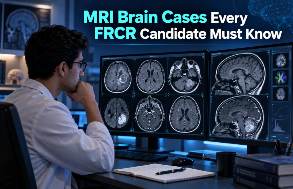
1. Acute Ischaemic Stroke
Key sequences: Diffusion-weighted imaging (DWI) and apparent diffusion coefficient (ADC) maps are critical.
Acute infarct shows restricted diffusion (bright on DWI, dark on ADC) within minutes to hours. FLAIR becomes hyperintense later. Look for vessel occlusion on MRA and susceptibility vessel sign on SWI.
Viva points: Describe the vascular territory, estimate age of infarct, comment on haemorrhagic transformation risk, and discuss eligibility for thrombolysis or thrombectomy. Always mention the need for CT or MR angiography if not already performed.
2. Brain Abscess vs Necrotic Tumour (GBM / Metastasis / Lymphoma)
This is a classic FRCR discriminator.
Abscess typically shows central restricted diffusion with thin, smooth ring enhancement and surrounding oedema. Necrotic tumours (especially GBM) show irregular, thick ring enhancement and variable diffusion in the solid components. Primary CNS lymphoma is usually solid, homogeneously enhancing, and restricts diffusion throughout.
Viva tip: Always comment on the pattern of diffusion restriction and enhancement. Mention the clinical context (fever, immunocompromise) and recommend urgent neurosurgical discussion and further imaging of the whole neuraxis if indicated.
3. Cerebellopontine Angle (CPA) Masses
The classic differential is vestibular schwannoma, meningioma, and epidermoid.
- Schwannoma: arises from the internal auditory canal, expands the porus acusticus, avid enhancement.
- Meningioma: broad dural base, dural tail, hyperostosis.
- Epidermoid: follows CSF on T1/T2 but restricts diffusion (bright on DWI).
Viva approach: Localise (intra- vs extra-axial), describe relationship to the IAC and cranial nerves, and offer the top three differentials with distinguishing features.
4. Pituitary Region Masses
Common cases include pituitary macroadenoma, craniopharyngioma, and Rathke’s cleft cyst.
Macroadenomas expand the sella, may invade the cavernous sinus, and show heterogeneous enhancement. Craniopharyngiomas often have cystic and calcified components (better seen on CT). Rathke’s cleft cysts are non-enhancing or thinly enhanced with characteristic location.
Always assess for optic chiasm compression and cavernous sinus involvement.
5. Ring-Enhancing Lesions
Use a structured differential:
MAGICAL DR or similar mnemonic (Metastasis, Abscess, Glioma/GBM, Infarct (subacute), Contusion, AIDS-related (toxoplasmosis/lymphoma), Demyelination, Radiation necrosis).
Emphasise number of lesions, location (grey-white junction vs deep), enhancement pattern, and diffusion characteristics. Clinical history (malignancy, infection, immunosuppression) is crucial.
6. Herpes Simplex Encephalitis
Classic involvement of the temporal and insular lobes, often bilateral but asymmetric, with restricted diffusion and variable haemorrhage on SWI/GRE. Sparing of the basal ganglia helps differentiate from MCA infarct.
Management point: Urgent antiviral therapy and neurology/infectious diseases input.
7. Multiple Sclerosis and Demyelination
Look for periventricular, callosal, and juxtacortical lesions (Dawson’s fingers), incomplete ring enhancement in active lesions, and involvement of the spinal cord or optic nerves. Differentiate from ADEM, NMO spectrum, and vascular disease.
8. Intracranial Haemorrhage and Vascular Malformations
Age blood products using T1/T2 signal (oxyhaemoglobin → deoxy → methaemoglobin → haemosiderin). Cavernomas show the classic “popcorn” appearance with blooming on SWI. Developmental venous anomalies have the Medusa-head appearance. AVMs show flow voids and feeding/draining vessels.
9. Hydrocephalus and Intracranial Hypotension
Distinguish obstructive vs communicating hydrocephalus. Spontaneous intracranial hypotension shows pachymeningeal enhancement, venous distension, subdural collections, and brain sagging.
10. Paediatric and Congenital Cases (High-Yield)
Posterior fossa tumours (medulloblastoma, ependymoma, pilocytic astrocytoma), neurocutaneous syndromes (NF1 optic pathway glioma, NF2 bilateral schwannomas, tuberous sclerosis), and malformations (Chiari, Dandy-Walker spectrum) appear regularly.
Frequently Asked Questions
1. Which MRI brain cases are most important for the FRCR 2B exam?
High-yield cases include acute ischaemic stroke, brain tumours, brain abscess, ring-enhancing lesions, cerebellopontine angle masses, pituitary lesions, multiple sclerosis, encephalitis, intracranial haemorrhage, vascular malformations, hydrocephalus, and paediatric brain abnormalities.
2. Why are MRI brain cases important for FRCR candidates?
MRI brain cases test several skills at once, including image interpretation, sequence recognition, lesion localisation, differential diagnosis, clinical reasoning, and appropriate management recommendations. These are important skills assessed during the FRCR 2B viva and reporting components.
3. Which MRI sequences should FRCR candidates know well?
Candidates should be comfortable interpreting T1, T2, FLAIR, DWI, ADC, SWI/GRE, post-contrast sequences, MRA, and MRV where appropriate. Understanding what each sequence demonstrates can help narrow the differential diagnosis quickly.
4. How should I approach an MRI brain case during an FRCR viva?
Start with a structured description. Identify the MRI sequences, localise the abnormality, determine whether the lesion is intra-axial or extra-axial, describe its signal and enhancement characteristics, look for diffusion restriction or haemorrhage, provide relevant differentials, and suggest appropriate further investigation or management.
MRI brain cases can appear challenging when multiple sequences, differential diagnoses, and management decisions need to be considered within a limited time.
For more FRCR exam tips, radiology case discussions, viva preparation, and high-yield revision content, visit and subscribe to our FRCRVIVACITY YouTube channel.
Watch. Learn. Practice. Prepare smarter for your FRCR exam.
👉 Subscribe to our YouTube channel for more FRCR learning videos and case-based revision.