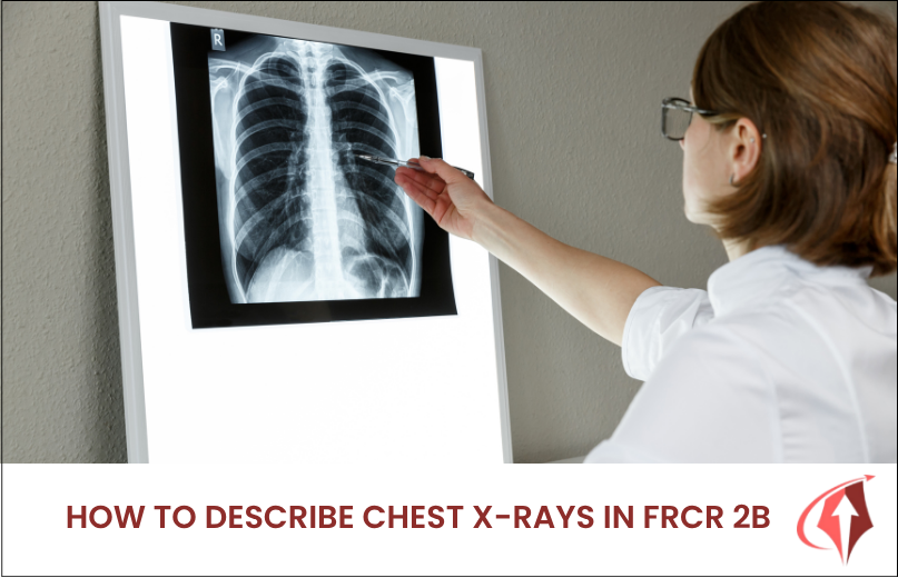
Chest radiographs remain one of the most frequently tested modalities in the Final FRCR 2B examination. Whether in the short-case reporting component (previously rapid reporting) or the viva, candidates are expected to describe findings clearly, interpret them accurately, and recommend safe next steps. A structured, consistent approach is essential – examiners value logical observation and clinical reasoning far more than lengthy descriptions.
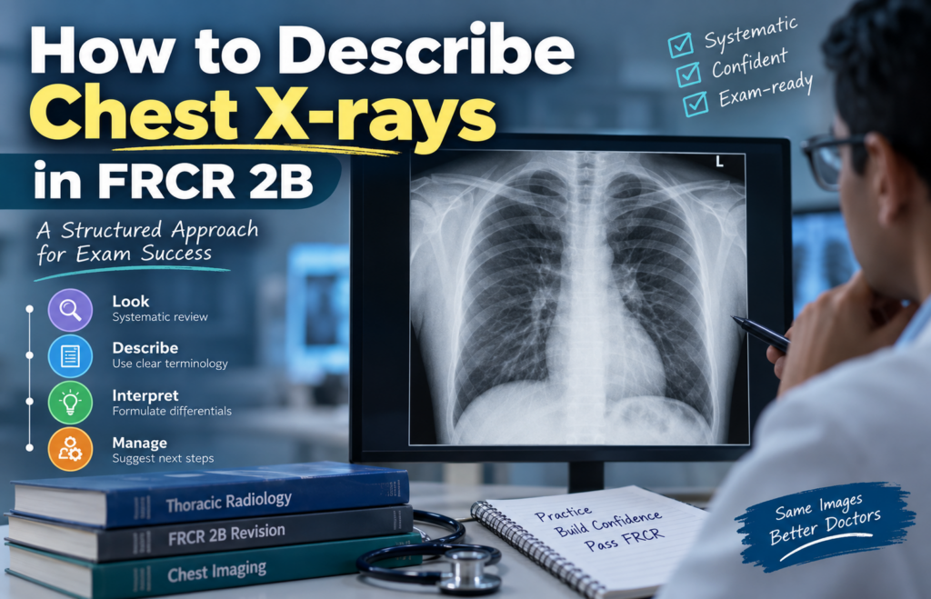
Why a Systematic Approach Matters
In the short-case reporting component, candidates must produce concise reports that include observations, a diagnosis (or ranked differential), and a management recommendation. In the viva, you will be expected to present findings fluently while demonstrating safety and clinical judgment. A rehearsed system prevents missed findings, reduces over-calling, and helps you stay calm under pressure.
Chest X-rays typically form 50 – 60% of the short-case set, so proficiency here directly impacts your score.
Step-by-Step Systematic Description
- Technical Assessment (Quick but Essential)
Start by noting:
- Projection (PA, AP, erect, supine, mobile)
- Side markers and patient details
- Rotation (clavicles equidistant from spinous processes)
- Inspiration (count anterior/posterior ribs)
- Exposure/penetration (vertebral bodies just visible behind the heart)
- Any artefacts or lines/tubes
Mention technical limitations only if they affect interpretation (e.g., “AP portable film with limited inspiration – heart size cannot be accurately assessed”).
- Initial Overview
Take a brief global look for obvious abnormalities (large pneumothorax, complete white-out, free gas under the diaphragm). Then proceed systematically so you do not miss secondary findings.
- Structured Review – A Practical Order
Many candidates use a modified ABCDE or anatomical approach:
- Airway / Mediastinum: Tracheal position, carina, mediastinal contours, aortic arch, aortopulmonary window.
- Bones and Soft Tissues: Ribs, clavicles, scapulae, spine, soft-tissue masses, surgical clips, breast shadows.
- Cardiac Silhouette: Size (cardiothoracic ratio on PA films), shape, borders.
- Hila: Position, size, density, and symmetry.
- Lungs and Pleura:
- Zones (upper, mid, lower) or lobes where possible
- Opacity type (consolidation, collapse, nodule, mass, interstitial)
- Distribution (focal, multifocal, bilateral, peripheral, central)
- Volume loss or expansion
- Pleural abnormalities (effusion, pneumothorax, thickening, plaques)
- Diaphragm and Below: Contour, free gas, gastric bubble, upper abdominal structures.
- Review Areas (high-yield for missed findings):
- Apices (Pancoast tumour, fibrosis, pneumothorax)
- Behind the heart (left lower lobe collapse, mass)
- Costophrenic angles
- Hila
- Below the diaphragm
- Soft tissues and bones at the periphery of the film
- Description of Abnormalities
Be precise and use radiological language:
- Location (zone or lobe, central/peripheral, unilateral/bilateral)
- Morphology (size, shape, margin, density, cavitation, calcification)
- Associated features (volume loss, shift of structures, air bronchograms, silhouette sign)
Avoid vague terms. Instead of “shadowing,” say “air-space opacification” or “reticular interstitial pattern.”
- Interpretation and Diagnosis
Link findings to a diagnosis or short differential. Rank differentials by likelihood and justify with imaging features and clinical information provided. For example:
- “Bilateral perihilar air-space opacification with Kerley B lines and bilateral pleural effusions – most consistent with acute pulmonary oedema. Differential includes infection or ARDS, less likely given the distribution and septal lines.”
- Management Recommendation
Always finish with a clear, safe next step:
- Further imaging (CT, previous comparison)
- Clinical correlation or urgent clinical review
- Referral pathway (e.g., respiratory team, MDT, interventional radiology)
- Follow-up radiograph after treatment
In short cases, keep the entire report concise – bullet points or short statements work well and are preferred by examiners.
Common High-Yield Patterns and How to Describe Them
- Lobar collapse: Describe volume loss, shift of fissures/mediastinum, and compensatory hyperinflation.
- Pneumothorax: Size estimate, presence of tension (mediastinal shift), and any underlying lung disease.
- Pleural effusion: Unilateral/bilateral, size, and any underlying mass or consolidation.
- Solitary pulmonary nodule/mass: Size, margins, location, calcification, and comparison with prior imaging if available.
- Diffuse opacities: Distinguish alveolar vs interstitial patterns and offer a focused differential (oedema, infection, haemorrhage, ARDS).
Viva-Specific Tips
- Begin with: “This is a frontal chest radiograph of an adult patient…”
- Describe positive findings first, then relevant negatives.
- Speak clearly and pause for the examiner.
- If you spot an obvious abnormality, describe it fully and then check review areas for additional findings.
- Never invent findings. If uncertain, say so and offer a sensible differential.
- Always consider the clinical history provided – it often narrows the diagnosis.
Practice Recommendations
- Develop and rehearse your own checklist until it becomes automatic.
- Practise under timed conditions for short cases.
- Present cases aloud regularly – this builds fluency for the viva.
- Review both classic and subtle cases, including paediatric films and lines/tubes.
- Learn common pitfalls: over-calling normal variants, missing review-area pathology, and failing to link findings to management.
Clear, structured description of chest X-rays is a core skill for FRCR 2B success. The more you practise presenting findings systematically, the more confident and accurate you will become.
Frequently Asked Questions
1. How long should a short-case chest X-ray report be?
Keep it concise – observations, diagnosis/differential, and management in a few clear statements or bullet points. Examiners prefer clarity over length.
2. Should I always mention technical quality?
Only if it significantly limits interpretation. Otherwise, move quickly to the findings.
3. What are the most commonly missed findings on chest X-rays in FRCR?
Apical lesions, left lower lobe collapse behind the heart, small pneumothoraces, free subdiaphragmatic gas, and bone lesions at the film edges.
4. Is it better to use zones or lobes when describing lung abnormalities?
Zones are safer on a single frontal film. Mention lobes only when the silhouette sign or fissure position allows confident localisation.
For realistic case-based practice that mirrors exam conditions, join the sessions on FRCR VIVACITY. Regular viva-style discussions help you refine observation, differential diagnosis, and communication skills across plain films and other modalities.
Explore the regular sessions and extensive video library on FRCR VIVACITY.
Join our Telegram community for regular updates, discussions, and exam-focused resources:
Telegram: https://t.me/frcr2bvivacity
Subscribe to our YouTube channel for more videos and updates!